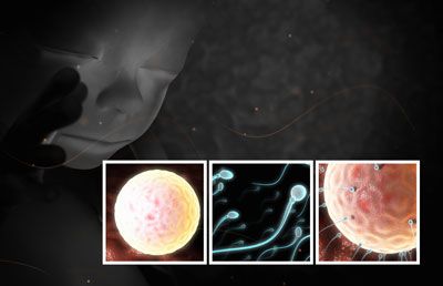
This illustration shows a close-up of the fetus in the womb. The smaller images show the sperm swimming, the unfertilized ovum in the fallopian tube, and the ovum surrounded by sperm. See how an embryo develops, next.
Advertisement

This series of images shows the development of the embryo. See how mothers and their doctors check up on the developing fetus, next.

A sonogram is used to confirm pregnancy in the first weeks of gestation. See another way sonograms are used, next.
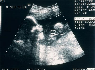
Sonograms can be used to look at the chambers of a fetus's heart as well as determine if the placenta is providing enough blood flow. See an embryo at six weeks in the next picture.
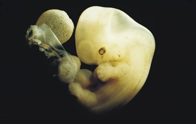
Human embryo at 6 weeks' gestation. Note the yolk sac, the umbilical stalk, and the developing notochord along the back that will become the spine. By the 8th week, the fetus is even more developed.
Advertisement
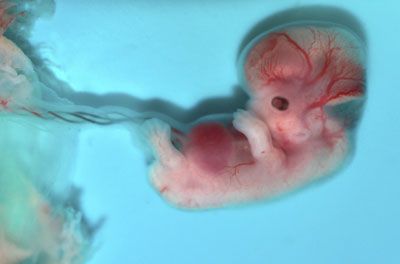
A human fetus during the 8th week of development. At this time the eye and the ear can be clearly seen, the limbs are developed and the notches between the fingers and toes are beginning to demarcate the individual digits. On to the second trimester now.
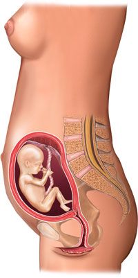
This stock medical illustration shows the fetus in the uterus in the fourth month (second trimester) of pregnancy. Next -- have you ever seen a fetus in 3D?

The Phillips 3D ultrasound showing an unborn child at Insight Radiology, Auckland New Zealand. Sonograms are still the most widely-used imaging technique, though. See that next.
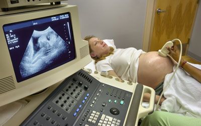
Ultrasound technicians use sonograms to measure the growth of a fetus and determine if there are any possible pre-existing conditions. Next, see what fundal height is.
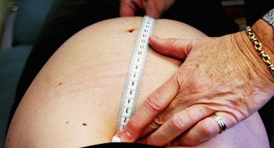
A midwife measures the fundal height of a pregnant woman during a routine check-up at Royal North Shore Hospital's Birth Centre June 7, 2006, in Sydney. Our last photo shows what makes it all worthwhile.
Advertisement

In the end, the fruits of a mother's labor end in a strong bond between mother and child. Learn all aboutHow Pregnancy Works.

